
Ultrasound Imaging of the Ear
Working to bring this technology into the clinic
Direct visualization of the auditory system is difficult due to the opacity of the tympanic membrane (TM) and/or the round window membrane. Even with cross-sectional imaging modalities, such as computed tomography or magnetic resonance imaging, diagnostically useful information can be difficult to obtain due to insufficient image resolution.
Ultrasonography is a technique not yet clinically applied to ear imaging, which could provide a great deal of useful information to otologists. The equipment size and safe usage considerations of ultrasound systems could allow such a method to be used at initial patient contact in the clinic.
Traditional ultrasonography, such as that used for fetal or cardiac imaging, operates at low frequency (typically 1-10 MHz) in order to obtain adequate penetration to reach the targeted anatomy. The use of low frequencies limits the axial and lateral resolution of such systems to a few hundred microns at best.
Recently, high-frequency ultrasound (HFUS) systems operating in the 30-60 MHz range have been developed by our research group and a few others. These systems are able to achieve much higher resolution on the order of tens of microns compared to traditional ultrasonography.
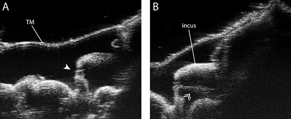
The images below represent examples of 50 MHz ultrasound images of a cadaveric middle ear generated in our facility. Some important anatomical features are clearly visible with this technology including the TM, the ossicles, and the incu-stapedial joint.
One of the primary research objectives of our group is to miniaturize the high resolution ultrasound imaging probes into endoscopic form factor so that we can bring this technology to the clinic for in vivo imaging and Doppler measurements.