Histology

Histology and Digital Pathology Core Facility (HistoCORE)

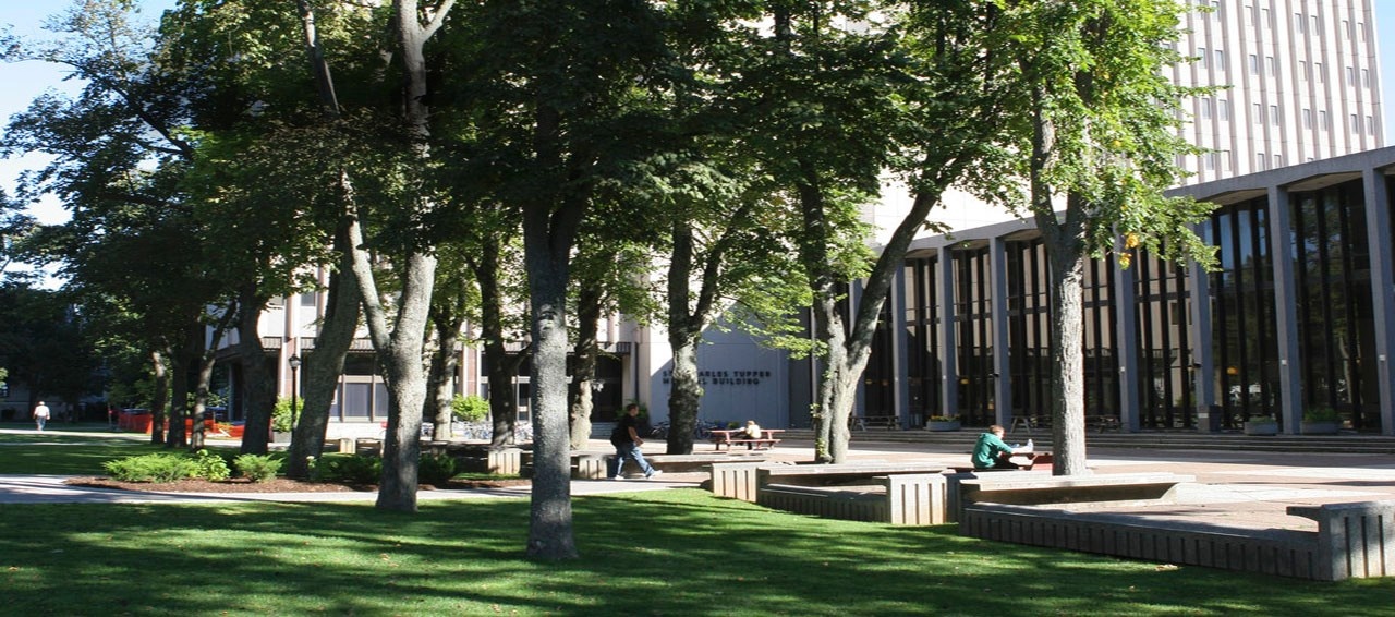
The Histology and Digital Pathology Core Facility (HistoCORE) is part of the Dalhousie Faculty of Medicine Centralized Operation of Research Equipment and Support program (CORES). HistoCORE is located in 13D Tupper Building and contains advanced instrumentation for a wide range of routine and advanced histology and digital pathology services, which include:

  1. Tissue processing and embedding: Fixed tissues are processed into paraffin blocks, for sectioning.
  2. Tissue sectioning: Paraffin embedded tissues are sectioned at 2 – 5 uM and mounted onto slides for downstream histological staining, immunohistochemistry/immunofluorescence or in situ hybridization studies. Frozen tissues are sectioned at 10-15 uM.
  3. Histology stains, such as H&E, PAS, Oil Red O and Masson’s Trichrome: Tissue slides are stained with histology dyes for observation of overall tissue architecture, as well as specific tissue features, such as collagen or mucopolysaccharides.
  4. Immunohistochemistry – single and duplex: Tissue slides are stained for one or two specific proteins of interest, using antibodies and chromogenic detection.
  5. Immunofluorescence: Tissue slides are stained for up to 6 specific proteins of interest, using antibodies and fluorescence detection, most commonly with OPAL fluorophores.
  6. In situ hybridization: Tissue slides are stained for a target RNA molecule, using chromogenic or fluorescence detection methods.
  7. Brightfield whole slide scanning: Instead of using a microscope to view and photograph regions of interest in a stained tissue, the whole tissue can be scanned at 20 or 40X then viewed and/or analyzed in its entirety.
  8. Fluorescence microscopy: Our Zeiss Axio Observer 7 microscope equipped with a Hamamatsu ORCA Fusion BT CMOS camera can be used to observe and photograph tissues stained with up to 7 fluorophores, with excellent resolution and sensitivity.
  9. Tissue microarray: Up to 96 tissue samples can be included on one slide with our TMA Master II microarrayer. Guided core selection from donor tissues is carried out, followed by insertion of the cores into a TMA block, according to a pre-established map.

Services upcoming will include:

  1. RNAScope: Tissues can be probed for up to 6 RNA targets simultaneously using a sensitive and specific staining method, and fluorescence detection. Alternatively, tissues can be probed for a combination of protein and RNA targets simultaneously, up to 6 in total.
  2. Fluorescence whole slide scanning with spectral unmixing: Tissues stained using multiplex immunofluorescence or RNAScope be scanned in their entirety using the PhenoImager HT, which will also separate overlapping fluorescent signals and remove autofluorescence, facilitating analysis of the entire tissue, rather than regions of interest only.
  3. Advanced digital analysis, such as quantitation, segmentation, phenotyping, hotspot and spatial mapping. VisioPharm Multiplex AI software can be customized to analyze multiple aspects of any tissue. In addition, analyses are speeded up by the batch processing capability of the software.

Equipment

  1. Pegasus Tissue Processor
  2. Arcadia Embedding Centre
  3. HistoCore AUTOCUT Microtome
  4. Cm1950 Cryostat
  5. Spectra ST+CV Autostainer
  6. Bond RX Automated IHC/ISH Multiplex Staining Platform
  7. TMA Master II
  8. Aperio XT Brightfield Whole Slide Scanner
  9. Axio Observer 7 Wide Field Fluorescence Microscope with ORCA Fusion BT Camera

Equipment coming soon will include:

  1. NanoZoomer S360 High Throughput Whole Slide Scanner
  2. PhenoImager HT Fluorescence Whole Slide Scanner
  3. VisioPharm Multiplex AI Analysis Software

FAQs

Do you offer customized project services?

We do. We are always happy to work with researchers on project design and method development. Please contact us at hiscore@dal.ca for more information.

How long will it take to complete my project?

We strive to complete routine services within 2 weeks, but timing does depend on the size and complexity of the project, as well as the queue for services. For a more precise estimate, please make an appointment to discuss, by emailing hiscore@dal.ca.

How do I submit my samples?

Please submit a HistoCORE requisition (available by emailing hiscore@dal.ca) via email, to set up a time to drop off your samples in 13D Tupper. Please list all samples individually in the requisition.

Do you have a price list for services?

We have a price list, which you can have emailed to you, by contacting us at hiscore@dal.ca . Since prices may change annually, please ensure that your price list is up to date.

Can I process my own samples?

No, samples must be left with HistoCORE staff at present. The only exception is cryostat services, for which we do offer self-service and full training.

Can I drop in any time?

We are always happy to engage with researchers, but we encourage them to make an appointment before coming to the HistoCORE, to avoid interruptions to colleagues who have meetings scheduled, or arriving when staff are not available for consultations.

How should I prepare my samples?

Please email us at hiscore@dal.ca for detailed instructions on how to prepare tissues and/or slides before submitting to the HistoCORE.

People

Lynn Thomas, Manager

Educated at Dalhousie University, with over 30 years of experience in histology, antibody validation, immunohistochemistry, microscopy and image analysis techniques, in addition to method development and project management.

Select publications (full list of 35 articles available by request):

  • MacDonald TM, Thomas LN, Gupta A, Barnes PJ, Too CK, 2020. Prolactin and androgen R1881 induce pro-survival carboxypeptidase-D and EDD E3 ligase in triple-negative and HER2+ breast cancer. Am J Cancer Res 10(5):1321-1343
  • Bharadwaj AG, Dahn ML, Liu RZ, Colp P, Thomas LN et al., 2020. S100A10 has a critical regulatory function in mammary tumor growth and metastasis. Cancers (Basel) 12(12):3673
  • MacDonald TM, Thomas LN, Daze E, Marignani P, Too CK, 2019. Prolactin-inducible EDD E3 ubiquitin ligase promotes TORC1 signalling, anti-apoptotic protein expression, and drug resistance in breast cancer cells. Am J Cancer Res 9(7):1484-1503
  • Thomas LN, Chedrawe ER, Barnes PJ, Too CKL, 2017. Prolactin/androgen-inducible carboxypeptidase-D increases with nitrotyrosine and Ki67 for breast cancer progression in vivo, and upregulates progression markers VEGF-C and RunX2 in vitro. Breast Cancer Res Treat 164(1):27-40
  • Thomas LN, Merrimen J, Bell DG, Rendon R, Goffin V, Too CK, 2014. Carboxypeptidase-D is elevated in prostate cancer and its anti-apoptotic activity is abolished by combined androgen and prolactin receptor targeting. Prostate 74(7):732-42
  • Lynn N Thomas and Roger Rittmaster, 2009. Effect of steroid 5alpha-reductases inhibitors on markers of tumor regression and proliferation in prostate cancer. Chapter in book: Androgen Action in Prostate Cancer; James Mohler, Donald Tindall eds; Springer Publishing
  • Thomas LN, Douglas RC, Lazier CB, Too CK, Rittmaster RS, Tindall DJ, 2008. Type 1 and type 2 5alpha-reductase expression in the development and progression of prostate cancer. Eur Urol 53(2):244-52 REVIEW

Contact us

Email: hiscore@dal.ca

Mail

Histology and Digital Pathology Core Facility
13D Tupper Building
Dalhousie University
5850 College St.
Halifax, NS B3H 4R2
Fees (Internal)

Service 

Unit (1)

Price 

     
Histological Services    
FFPE    
Biobank Clinical Data/Sample Access and Retrieval Fees  Patient $30/patient case
Paraffin Wax Processing, Embedding   Block  $10.50/block 
Paraffin Wax Processing, Embedding, Cut 5um Block/1slide  $16.50/block/1slide 
Cut additional slides FFPE Slide  $6 

Human Tissue Control Slides FFPE 


Slide 


$15 
HistoGel-Gelatin Cell pellet creation- without embedding  Tube $80 
Deparaffinize

Slide 


$3


Cut sections-sterile (2x20um for DNA extraction) 


Tube 


$10 


H&E Staining + Mounting 


Slide 


$3.50 


Counterstain + Mounting 


Slide  


$3.50 


Hoechst Stain + Mounting 


Slide 


$10 


Microtome (Self Service)  


Hour 


$20 


Microtome (Initial Training-Self Service)  


Hour 


$80 


Frozen 


Cryostat, Tissue Prep. (10um section) 


Block/1slide 


$25/block/1slide 


Cryostat, Cut additional slides (10um)  


Slide 


$10 


Cold Acetone Fixation for frozen sections 


Slide 


$4 


Cryostat (Self Service) 


Hour 


$30 


Cryostat (Initial Training-Self Service)  


Hour 


$80 


Other Additional Services 


Antigen Retrieval- De clocker 


Run 


$8 


Citrate Buffer pH:6.1 


Run 


$7/rack (max 3 racks/run)  


Special Stains 


Masson’s Trichrome 


Slide 


$40 


PAS 


Slide 


$15 


Oil red O  


Slide 


$15 


Verhoff Van Gieson 


Slide 


$20 


Alcian Blue 


Slide 


$15 


TMA  


Set-up fee (up to 48 Cores, higher charges will apply for greater core number) 


Up to 48 Cores 


$450 


TMA Construction 


Per Core 


$7 


TMA Sectioning  


Slide 


$12 


Digital Slide Scanning  


Whole Slide Scanning BF (20X) 


Slide 


$8 


Whole Slide Scanning BF (40X) 


Slide 


$11 


Whole Slide Scanning F (20X) 


Slide 


$20 


Whole Slide Scanning F (40X) 


Slide 


$30 


+ Whole Slide Scanning Tech Time (estimated at 1hr/20 slides) 


Hour 


$80 


Digital Image Storage (Memory USB) 32 - 64GB 


$10 - $30 


Digital Pathology Analysis  


inForm Software: Deconvolution-Spectral Unmixing-Removal of Autofluorescence- Quantitation 


Hour + 


$80 


VisioPharm Multiplex AI: Tissue and Cell Segmentation-Automated Phenotyping- On Tissue Spatial Mapping- 


Nearest Neighbor Analysis-Crosstalk Analysis-Component Reporting- Heat Maps- Hot Spot Analysis-Rare Event Detection- Images Co-Registration and Colocalization and others (Hours depends on tissue and staining complexity) 


Hour + 


$80 


+ VisioPharm- Single Plex 


Slide 


$15 


+ VisioPharm-Duplex-3Plex 


Slide 


$30 


+ VisioPharm-4-5Plex 


Slide 


$45 


+ VisioPharm-6 Plex 


Slide 


$60 


IHC-ISH Staining Services-Leica BOND RX- Single and Multiplex HRP and Fluorescence (OPAL dyes) 


Primary antibodies supplied by Researcher unless specified -  


Inquire about HistoCORE list of primary Ab Panels and PhenoCode Spatial Signature Panels  


Primary Antibody Optimization (HRP) 


Slide 


$60 


Platform - Leica Bond RX (HRP) 


Primary 


$10.50 


IHC -HRP (primary supplied by researcher) (1Unit) 


Slide 


$60 


Duplex IHC-HRP (2Units)                                            


Slide 


$120 


3-Plex IHC-HRP (2Units)                                            


Slide 


$120 


IF-Fluorescence-OPAL dyes 


Primary Antibody Optimization (includes 1 IF-OPAL) 


Slide 


$75 


Platform Leica Bond RX (IF) 


1 Primary Antibody 


$10.50 


IF- OPAL (without optimization) (1Unit) 


Slide 


$75 


OPAL Optimization A: Establish fluorochrome Ab combinations depending on Plex number Tech Time 


Hour 


$80 


OPAL Optimization B1: Establish Ab sequence depending on Plex number) Tech Time 


Hour  


$80 


OPAL Optimization B2: Sequence testing (3-Plex+Neg Ctr-2 slides) Total/Procedure 


Unit 


$300 


OPAL Optimization B3: Sequence testing (4-5 Plex+Neg Ctr-3 Slides) Total/Procedure 


Unit 


$675 


OPAL Optimization B4: Sequence testing (6 Plex+Neg Ctr-4 Slides) Total/Procedure 


Unit 


$1200 


Duplex IF (2Units) 


Slide 


$150 


3-Plex IF (2Units) 


Slide 


$150 


4-5 Plex IF (3Units) 


Slide 


$225 


6 Plex IF (4Units) 


Slide 


$300 


Histological services Formalin-Fixed Paraffin-Embedded (FFPE)

Service 

Unit (1)

Price

Biobank clinical data/Sample access and retrieval fees

Patient 

$30/patient case

Paraffin wax processing, embedding  

Block

$10.50/block

Paraffin wax processing, embedding, cut 5um

Block/1 slide  

$16.50/block/1 slide

Cut additional slides FFPE

Slide

$6 

Human tissue control slides FFPE

Slide

$15

HistoGel-gelatin cell pellet creation without embedding 

Tube

$80

Deparaffinize 

Slide 

$3.00

Cut sections-sterile (2x20um for DNA extraction)

Tube

$10 

H&E staining and mounting

Slide

$3.50

Counterstain and mounting 

Slide 

$3.50

Hoechst stain and mounting

Slide 

$10

Microtome (self-service) 

Hour

$20 

Microtome (initial training self-service) 

Hour

$80