How coronaviruses quietly shut down the body’s defenses
» Go to news mainDalhousie‑led study reveals how a key viral protein disrupts cells’ internal delivery system and helps virus spread
")
Inside infected cells, coronaviruses are doing more than building copies of themselves—they’re quietly shutting down the body’s ability to fight back.
A trainee-led study from Faculty of Medicine researchers at Dalhousie, published in PLOS Pathogens, has uncovered a previously unknown function of the coronavirus Membrane protein, also known as “M.” The discovery helps explain why coronaviruses are so effective at evading the immune system—and points to new possibilities for treatment.
Long understood as a structural component that helps assemble new virus particles, the team discovered that M also disrupts the cell’s internal delivery system—the network responsible for moving critical immune signals to where they’re needed. Recent studies have identified experimental antiviral drugs that target the M protein, and this work suggests those drugs could have a dual effect: preventing the virus from assembling while also allowing the body’s natural antiviral defenses to function again.
In healthy cells, this internal system acts like a postal service, transporting proteins that help coordinate immune responses. But the study found that when the virus produces the M protein, it creates a traffic jam, preventing those signals from reaching their destination.
“We found that the M protein isn’t just structural—it influences how our cells respond during infection,” says Dr. Taylor Caddell, who led this work during her PhD studies along with research associate Dr. Eric Pringle, and supervisor Dr. Craig McCormick.
“It’s more than a building block for the virus—it undermines our response to infection.”
That disruption gives the virus a powerful advantage. When immune signals can’t move through the cell, the body’s ability to respond to infection is weakened. At the same time, the virus continues to replicate and spread.
What makes this response especially striking, researchers say, is its efficiency. By interfering with the host cell delivery system, the virus blocks key defenses without disrupting its own escape route. Newly formed coronaviruses use a different pathway to leave the cell, meaning they aren’t affected by the same traffic jam they create.
“This is where it gets really interesting,” says Dr. Pringle. “The virus sets up shop and, in doing so, blocks all of the normal cellular proteins that need to move through that pathway.”
Importantly, the M protein is highly conserved across coronaviruses, meaning it changes very little from one virus to another. According to Dr. McCormick, that opens the door to broader applications.
“When you’re dealing with a viral protein like M that has a conserved, fundamental role, it becomes very difficult for the virus to overcome challenges like antiviral drugs through mutation and evolution. Theoretically, this should mean that antiviral drugs that target M should not be so easily defeated by rapid viral evolution.”
From a pandemic preparedness perspective, that possibility is significant. Approaches that focus on shared, essential viral mechanisms could lead to treatments that are effective against multiple coronavirus threats, including future ones.
“If you can interfere with these fundamental processes, you’re not just working against one virus—you’re creating opportunities to target an entire family of related viruses with pandemic potential,” says Dr. Caddell. “That opens up the possibility of having tools ready for outbreaks that haven’t happened yet.”
Groundwork for future breakthroughs
The study also highlights the importance of fundamental research. While vaccines and treatments often grab headlines, discoveries like this—focused on understanding how viruses work at a basic level—lay the groundwork for future breakthroughs.
This includes a growing area of research into how viruses manipulate cellular metabolism. At Dalhousie, this work is strengthened by interdisciplinary collaboration, drawing on expertise from fields such as diabetes and metabolic disorders to better understand how viruses take over and alter cellular metabolism for their own purposes.
“This is where it’s really exciting to be part of a broader Faculty of Medicine community,” says Dr. McCormick. “We have colleagues working on metabolism and related diseases who already have the tools and knowledge we need to start answering these questions.”
Looking ahead
Looking ahead, the researchers are eager to build on these findings—supported by new tools that allow them to explore these questions in much greater detail.
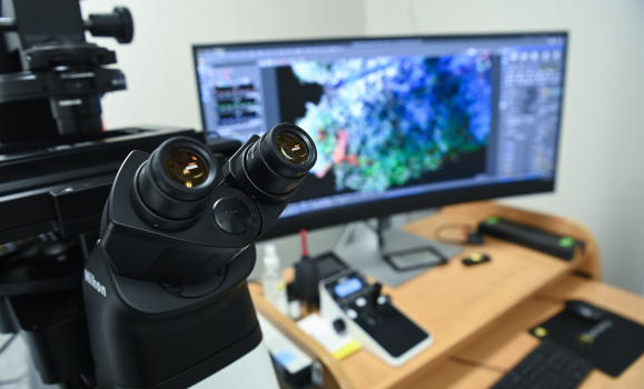
At the centre of this next phase is an advanced microscope that enables scientists to observe cellular processes in real time. Unlike earlier technologies that captured only static images, the system records fast-moving events inside cells, offering a far clearer picture of how viral proteins reshape their environment.
“The new microscope allows us to capture events as they happen, in much higher resolution,” says Dr. Caddell. “That means we can actually watch processes unfold in real time, which is much more informative.”
That capability could significantly accelerate discovery—helping researchers move beyond what happens to understanding how and why it happens. For Dr. Pringle, the advantage is both speed and precision.
“It’s faster and can capture much smaller changes. A lot happens inside cells very quickly, and without the right tools, you only see the start and end—not how things actually happen.”
At the same time, these advances bring new challenges. The volume of data generated by modern imaging technologies is enormous, requiring new approaches to analysis and new expertise to manage and make sense of it.
Support from CORES
Enabling this work is a broader ecosystem of expertise and shared resources. Dalhousie’s Centralized Operations of Research Equipment and Supports (CORES) facilities play a critical role in managing sophisticated equipment and ensuring researchers can make the most of it.
“CORES staff share their exceptional expertise with Dalhousie researchers, providing training and and helping design experiments that make the most of high-tech shared equipment,” says Dr. McCormick. “Experts like Dr. Gerard Gaspard, manager of the Cellular & Molecular Digital Imaging facility, are key to our success.”
That kind of support ensures researchers have not only the tools, but the expertise needed to push questions further and uncover new insights.
Inside infected cells, viral processes unfold quietly and out of view. With advanced tools and a collaborative research community, however, Drs. Caddell, Pringle, and McCormick are bringing them into focus—revealing not just how viruses operate, but how they might be stopped.
Recent News
- Manufacturing the future of life‑saving medicine in Nova Scotia
- Celebrating this year’s Excellence in Leadership award recipients
- The 2026 annual Faculty Awards
- New Dalhousie study tracks growing administrative burden on family physicians
- Dalhousie‑led study reveals how a key viral protein disrupts cells’ internal delivery system and helps virus spread
- First cohort of IPMP students graduate from Medical Sciences Program
- A First look at medicine beyond the city with Dr. Sebastian Copp
- Dr. Andrew Harding receives Class of 2026 C.B. Stewart Medal